My Personal Experience With Microscopes
The Letter "e" under a Microscope- 40x
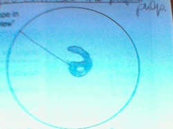
Ever looked at letters through a microscope? Well the outcome it funky because the letter is flipped upside-down, backward, and HUGE!! compared to its size with the naked eye.
-->why? light reflects weird and your brain actually has to flip images for you. but this time it can't- because you are looking through 2 lens (eyepiece and objective lens).
Also, another cool thing is that up close the letters' edges are not solid and straight, and you can see the paper's pulp.
-->why? light reflects weird and your brain actually has to flip images for you. but this time it can't- because you are looking through 2 lens (eyepiece and objective lens).
Also, another cool thing is that up close the letters' edges are not solid and straight, and you can see the paper's pulp.
How to determine how greatly magnified the view is:
eyepiece (10X) X Objective lens (ex. 10x)= total (100x)
Yes, its that simple.
Moving the Slide
One thing to be noticed when moving the slide is that it moves in the opposite direction (for example, if I move it to the right the letter moves to the left)
--> why does it move like this? because of the light reflection and having 2 lenses that flip the image.
--> why does it move like this? because of the light reflection and having 2 lenses that flip the image.
Looking at Hair Samples: Dark and Light
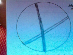
This is a sketch of the view of two human hairs via a traditional classroom light microscope (40x). The dark colored hair is actually on top of the light colored hair, although it is hard to see.
*Note: the diameter of the field is 1500 micrometers
When you look with a higher power objective lens the field of view is divided by 10 micrometers
These two hairs cross and I cannot see both hairs sharply at the same focus level. I used the fine adjustment knob to determine which hair(the dark brown one) is crossed over the other. It was hard because the microscope can see through different dimensions, thus the reason why the whole object was not in focus is because the microscope is unable to focus in on 2 different dimensions.
The Hairs under High Power (400 x)
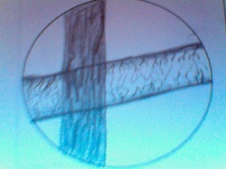
This is a sketch of the view of two human hairs via a traditional classroom light microscope. The dark colored hair is actually on top of the light colored hair, although it is hard to see.
-->why is is hard to tell which overlaps? well because microscopes can see to 2 different dimensions
How to calculate the diameter in micrometers of the high power field of view?
magnification # of high power objective/magnification # of scanning objective = (ratio) A
diameter of scanning power field of view/ A= diameter of the High Power Field of View
So...
the diameter in micrometers of the high power field of view is 150 micrometers
TAKING A CLOSER LOOK AT CELLS
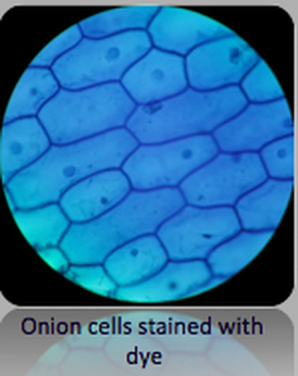
Onion Cells
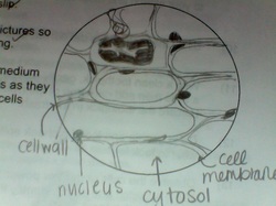
Recently, I took a look at onion cells under a national light microscope.
<-- A sketch of onion cells at high power (40x)
These cells appear to have 3 dimensions
Note: when looking with one eye, the object has 2 dimensions, but when looking at something with both eyes open, it appears to have 3 dimensions
Also, these onion cells have been labeled for your convenience.
Confused about what each part of the cell is?
- nucleus: organelle that stores the cell's genetic information (only in eukaryotes)
- cell wall: a special feature belonging just to plant cells. They are located outside of the cell membrane and are less flexible.
- cytosol: the gel-like liquid that is inside of the cell. In this substance, all of the cell's functions take place and all organelles are located in this. its part of the cytoplasm.
- cell membrane: all cells have a cell membrane that circle around the cell and are the cell's boundary. Made of phospholipids.
I have also looked at Elodea Cytoplasmic Streaming (check out the cool video below)
--> what is Cytoplasmic Streaming? Well "Cytoplasmic streaming" is defined as the movement of the fluid substance (cytoplasm) within a plant or animal cell(and in this case, in a 100 micrometer long elodea cell. ). This is the mechanism for the transport of nutrients.
-->But why can we see this streaming? Well, evaporation of some of the water makes it possible to see chloroplasts in motion inside of the cytoplasm.
Elodea Cell
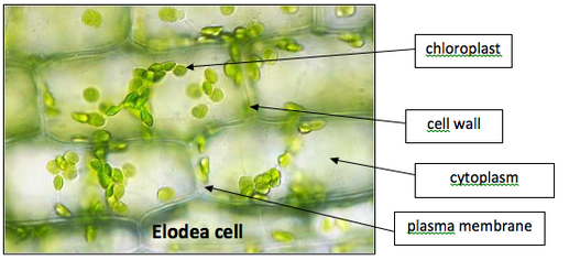
Here a high-resolution picture of a 100 micrometer long elodea cell, labeled.
Elodea Cytoplasmic Streaming
I have also looked at Elodea Cytoplasmic Streaming (check out the cool video below)
--> what is Cytoplasmic Streaming? Well "Cytoplasmic streaming" is defined as the movement of the fluid substance (cytoplasm) within a plant or animal cell (and in this case, in a 100 micrometer long elodea cell. ). This is the mechanism for the transport of nutrients.
-->But why can we see this streaming? Well, evaporation of some of the water makes it possible to see chloroplasts in motion inside of the cytoplasm.
One important piece of information to keep in mind is that this elodea cell is living?
--> Is there any evidence that is cell is living?
First of all, for something to be classified as "living" it must have these
8 LIFE FUNCTIONS.
R- Respiration
R- Regulation
R- Reproduction
E- Excretion
G- Growth
N- Nutrition
T- Transport
S- Synthesis
we know that this cell is living because we see the chloroplasts in motion inside of the cell. If the cell wasn't alive the chloroplasts wouldn't be alive either.
Dyes and Microscopes
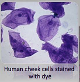
Methylene blue solution was used to help see check cells better but -->why do biologists use stains to study cells?
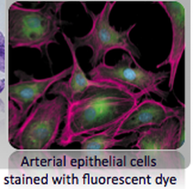
Because most cells are transparent, making them difficult to see without staining them with dyes!
Here are some pictures of cells that have been stained, for scientists (as ourselves) to see them better:
Because most cells are transparent, making them difficult to see without staining them with dyes!
Here are some pictures of cells that have been stained, for scientists (as ourselves) to see them better:
Want more information on microscopes and their uses?
Click the button below:
Click the button below:
Common Organelles in plant and animal cells

Here is a chart of common organelles that I discovered in both plant and animal cells.
Really, the only difference that I could see between these two types of cells was that plant cells have an (additional) cell wall.
But of course, there plenty of other differences between plant and animal cells…
Really, the only difference that I could see between these two types of cells was that plant cells have an (additional) cell wall.
But of course, there plenty of other differences between plant and animal cells…
Complete chart of Similarities and Differences in Plant and Animal Cells

Here is a fantastic chart taken from the book Miller and Levine: Biology
©2012 by AyushiSinhaMicroscopy
